

- 10.Liver
- ★(12)Hepatocellular carcinoma

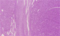
Microscopic finding (HE stain, low power view): On the right side is solid type hepatocellular carcinoma. On the left side is the background cirrhotic liver. The two areas are separated by fibrous capsule.
Click the image to see the enlarged image.