

- 10.Liver
- ★(1)Chronic viral hepatitis
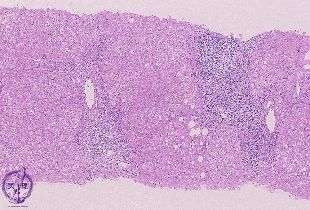
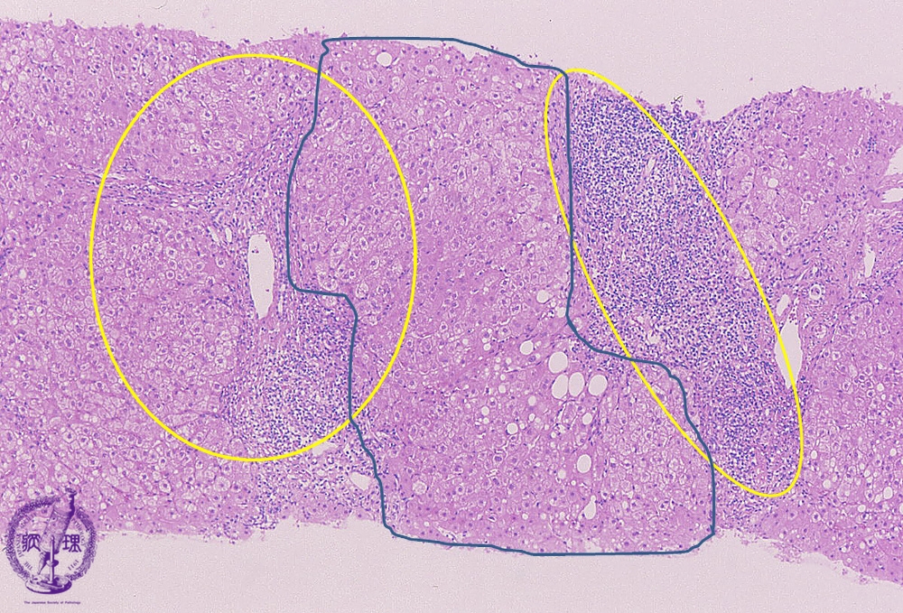
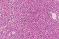
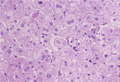
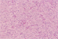
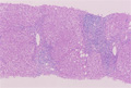
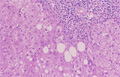
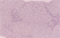
Microscopic image (HE stain, low power view): There is a dense lymphocytic infiltrate with corresponding moderate to marked portal expansion (yellow circle). Fibrosis (blue line) is also seen, and results in disruption of the lobular architecture with segregation of lobules which imparts a nodular appearance in some areas (blue area).
Click the image to see the enlarged image.