

- 10.Liver
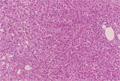
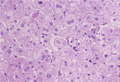
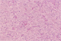
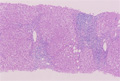
- ★(1)Acute viral hepatitis
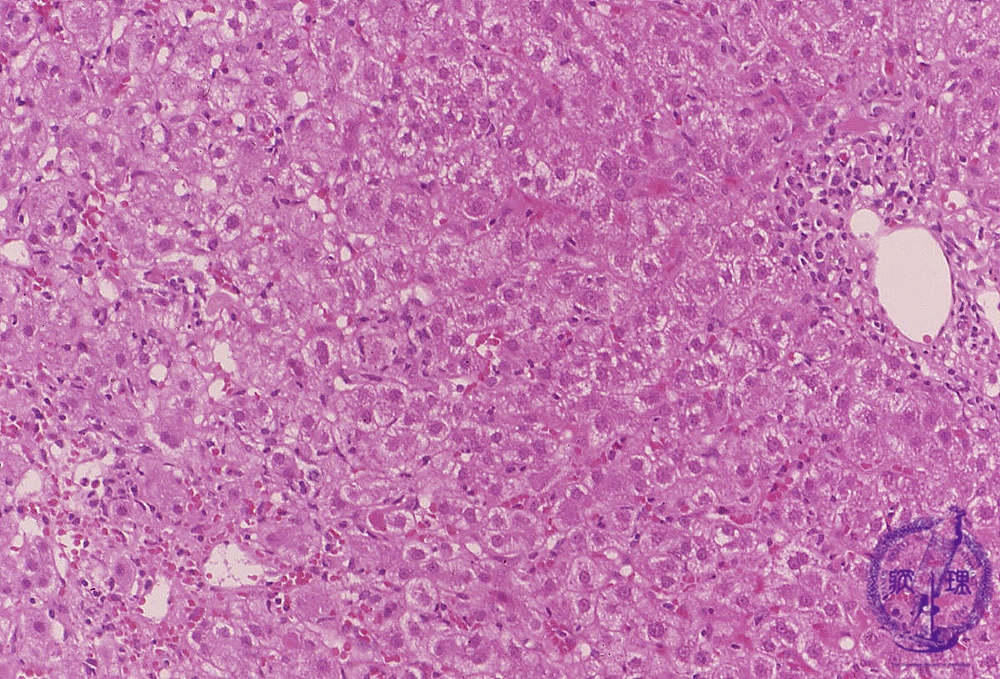
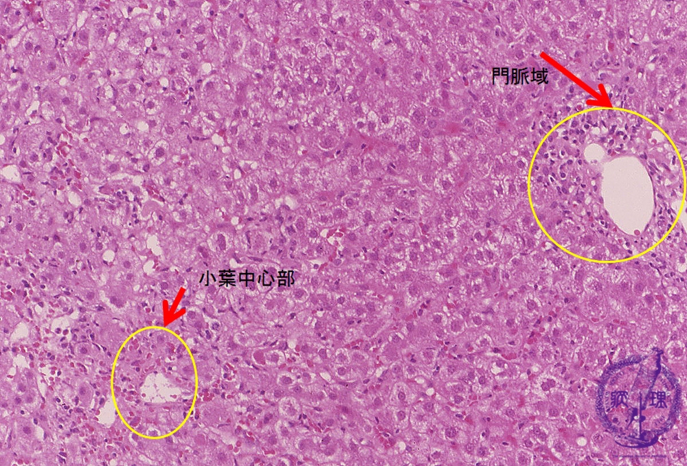
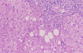
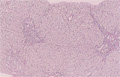
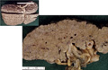
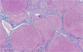
Microscopic finding (HE stain): There is diffuse involvement of lobules throuought the liver. Hepatocellular swelling, focal necrosis and inflammation are especially noticeable in the centrilobular area (image, lower left). Additionally, there is a periportal inflammation (image, upper right).
Click the image to see the enlarged image.