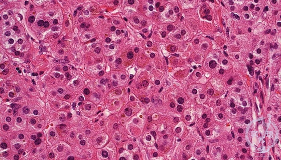
Microscopic findings (HE stain, high power view). Compact cells, with relatively high N/C ratio, demonstrated eosinophilic cytoplasm. The cells have a brownish hue due to the presence of lipofuscin granules.
Click the image to see the enlarged image.