

- 23.Cytology
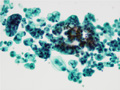
- (3)Cytology of peritoneal effusion( signet ring cell carcinoma of stomach)

(left: Papanicolaou stain, right: PAS stain)The tumor cells, enlarged nuclei with dense chromatin, displaced to the periphery by large mucous vacuole. In the normal condition, we could not see such atypical glandular epithelial cell in effusion.The cells contained PAS positive mucin. The mucin displaced the enlarged nuclei to the periphery. The cell border is demarcated.
Click the image to see the enlarged image.