

- 20.Skin
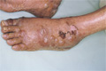
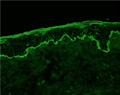
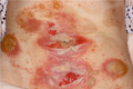
- ★(2)Vesiculobullous disease (Pemphigus vulgaris)
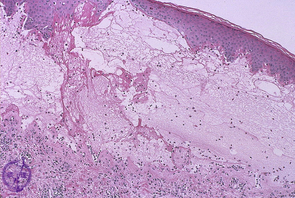
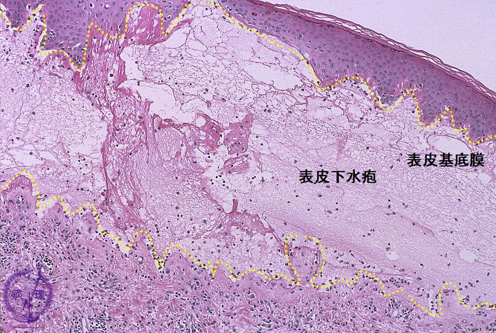
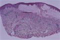
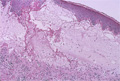
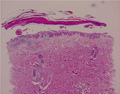
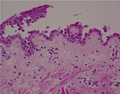
Microscopic image (Bullous pemphigoid): Fibrin deposition and numerous eosinophils are seen in the blister cavity. Eosinophil infiltration is also seen in the superficial dermis.
Click the image to see the enlarged image.