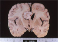


- 17.Nervous system
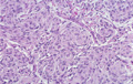
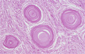
- ★(9)Meningioma
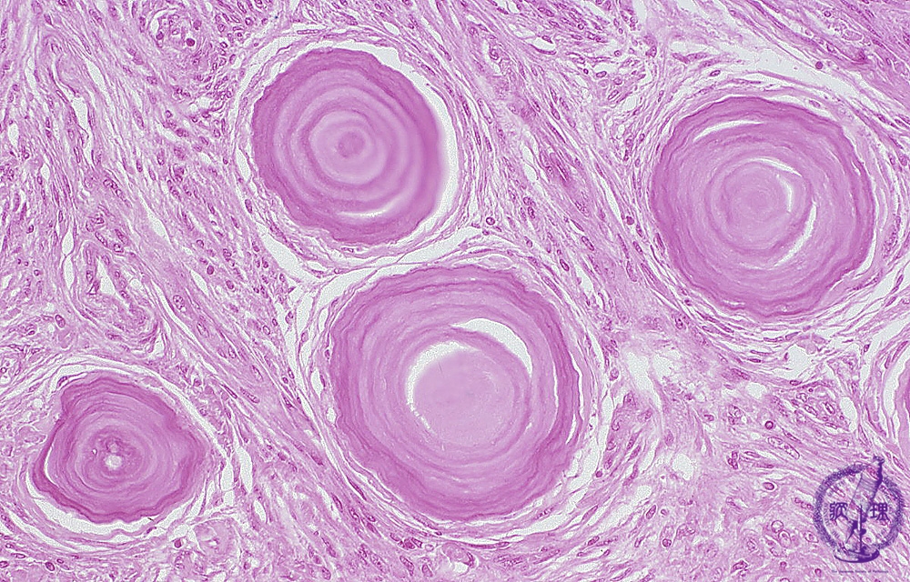
Microscopic findings (H.E. middle magnification): Psammomatous meningioma showed a proliferation of elongated tumor cells with fibrous bundles. Lamellar calcified bodies, namely psammoma bodies, were seen in a scattered fashion.
Click the image to see the enlarged image.