

- 17.Nervous system
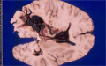
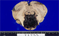
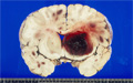
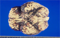
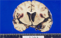
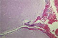
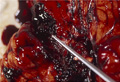
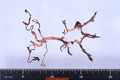
- ★(1)Cerebral hemorrhage(Berry aneurysm)
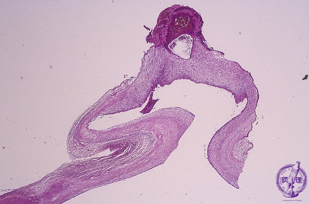
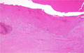
Microscopic findings (H.E. low magnification): Cystic dilatation and thinness of the brain arterial wall is visible. The wall was ruptured in part and the defect of tunica media and fibrin thrombi identified in the wall.
Click the image to see the enlarged image.