

- 12.Kidney and Urinary System
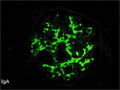
- ★(4)IgA nephropathy


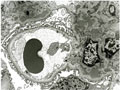
Microscopic findings (immunofluorescent microscopy, high power view): IgA deposits are demonstrated in the mesangium (red arrows).
Click the image to see the enlarged image.
Microscopic findings (immunofluorescent microscopy, high power view): IgA deposits are demonstrated in the mesangium (red arrows).
Click the image to see the enlarged image.