

- 12.Kidney and Urinary System
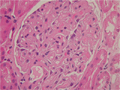
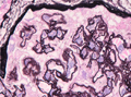
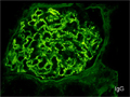
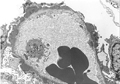
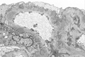
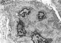
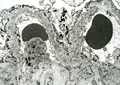
- ★(2)Membranous nephropathy
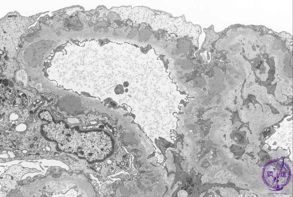
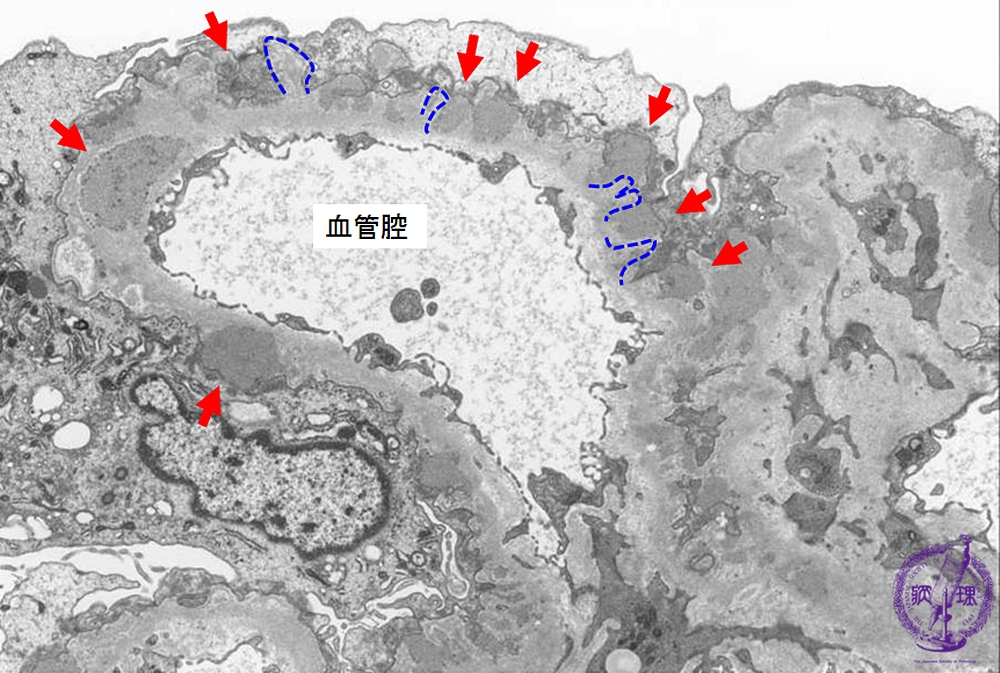
Electron microscopic findings: Stage II (focally Stage III). The number and size of electron dense subepithelial deposits (red arrows) are increased and spikes are seen forming between deposits (dotted blue line).
Click the image to see the enlarged image.