

- 11.Gallbladder, Pancreas
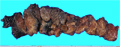
- ★(4)Acute hemorrhagic necrotizing pancreatitis, pancreatic pseudocyst
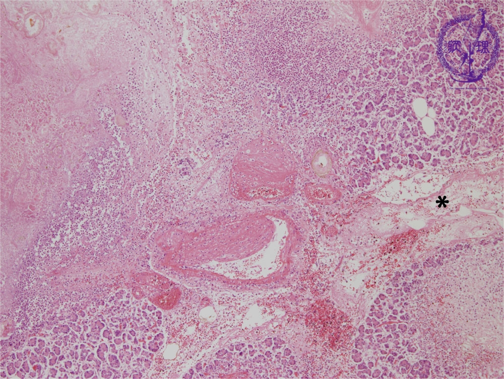
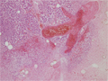
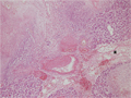
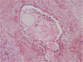
Microscopic image(HE stain, low power):There is destruction of interlobular blood vessels with pancreatic parenchymal necrosis. The majority of inflammation is seen within the pancreatic parenchyma.The interlobular space (*) shows edema and lymphectasia.
Click the image to see the enlarged image.