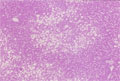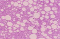


- 10.Liver
- (8)Fatty liver
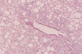
Microscopic image (silver stain, high power): Centrilobular hepatocytes demonstrate pericellular fibrosis and central vein walls are thickened. There is macrosteatosis.
Click the image to see the enlarged image.
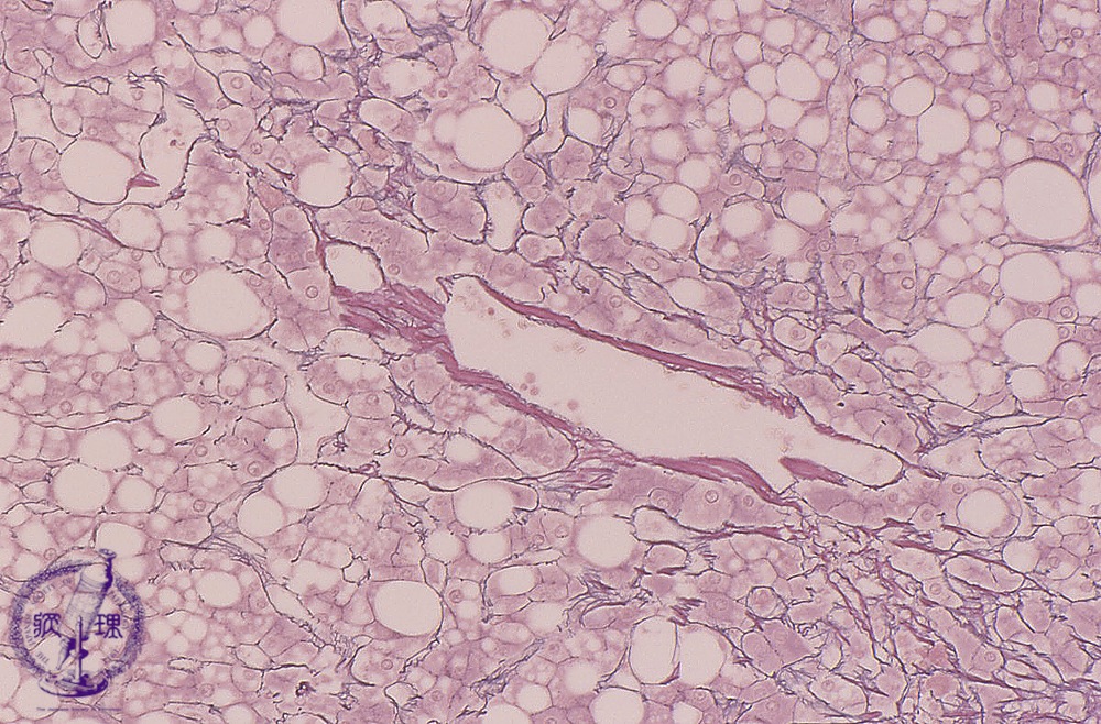
Microscopic image (silver stain, high power): Centrilobular hepatocytes demonstrate pericellular fibrosis and central vein walls are thickened. There is macrosteatosis.
Click the image to see the enlarged image.