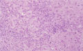


- 10.Liver
- (5)Wilson's disease
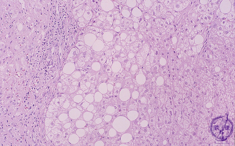
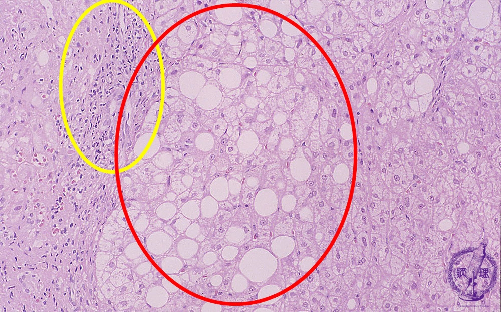
Microscopic image (HE stain, intermediate power): Various lesions are observed in the figure. On the left side are infiltrating lymphocytes (yellow circle) and at the center are steatosis and ballooning hepatocytes (red circle). To the right side, there do not appear to be any immediately identifiable abnormalities.
Click the image to see the enlarged image.