

- 7.Oral, Salivary gland
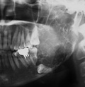
- (5)Odontogenic tumors(★Ameloblastoma)


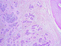
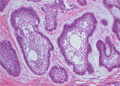
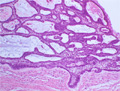
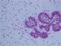
Microscopic finding (HE stain, intermediate-power view):The individual nests consist of two components. In the center are loosely arranged spindle or angular cells resembling stellate reticulum of the enamel organ. In the outer layer are columnar or cuboidal cells with nuclear palisading (arrows). Microcysts are formed within the nests (parenchymal cyst*) (follicular type)
Click the image to see the enlarged image.