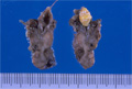


- 7.Oral, Salivary gland
- (1)Sialolithiasis
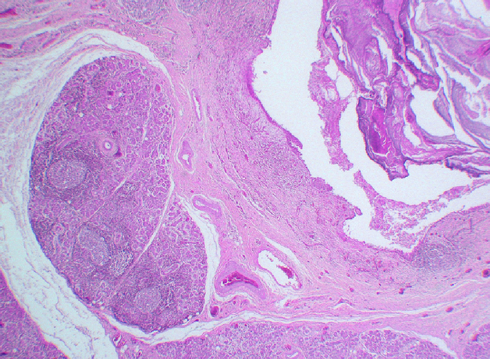
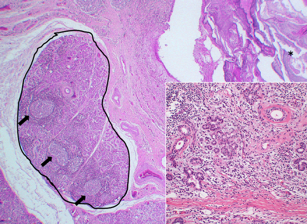
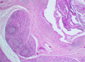
Microscopic finding (HE stain, low-power view). A sialolith (*), with concentric laminations, is seen lodged inside a markedly dilated duct. There is pronounced chronic interstitial inflammation of salivary gland lobules (○) with lymphoid follicle formation (arrows). Intra and perilobular fibrosis are characteristic findings in chronic sclerosing sialadenitis. There is acinar atrophy and destruction with ductal dilatation (lower right).
Click the image to see the enlarged image.