

- 3.Cardiovascular system
- (12)Varicose veins
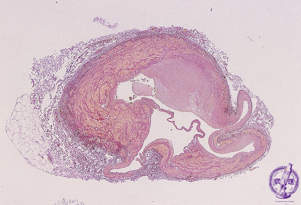
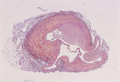
Microscopic image (EVG, low power view): Histopathology of varicose vein. There is venous wall thickening, fibrosis and focal aneurysm composed of vascular wall dilatation.
Click the image to see the enlarged image.