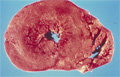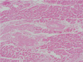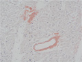


- 3.Cardiovascular system
- (8)Cardiac amyloidosis
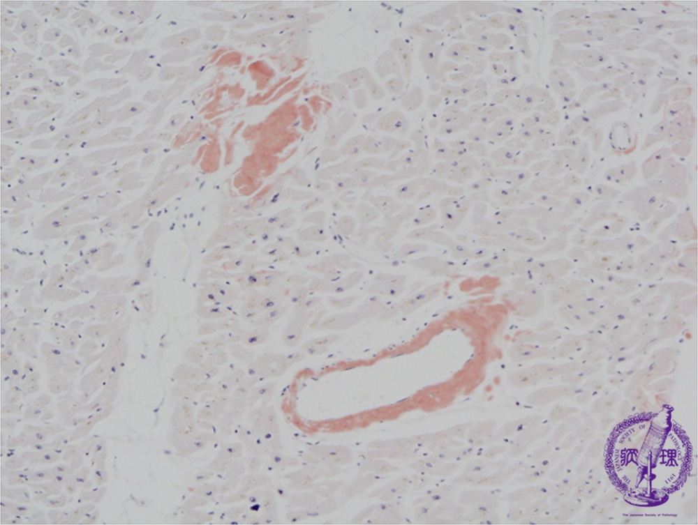
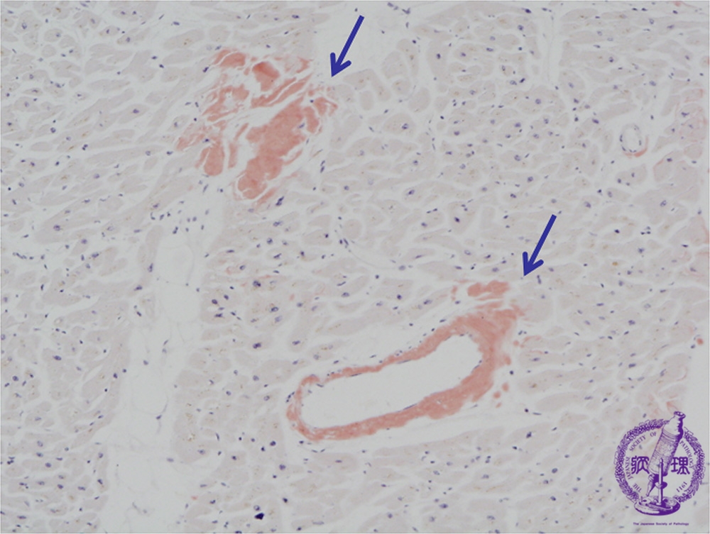
Microscopic image (Congo-red stain, low power view): There are small deposits of amorphous substance on HE staining composed of amyloid as demonstrated by Congo red stain (arrow). Amyloid deposition is confirmed by polarized light microscopy, which shows focal apple green birefringence.
Click the image to see the enlarged image.