

- 2.Lymph node
- (2)Tuberculous lymph nodes
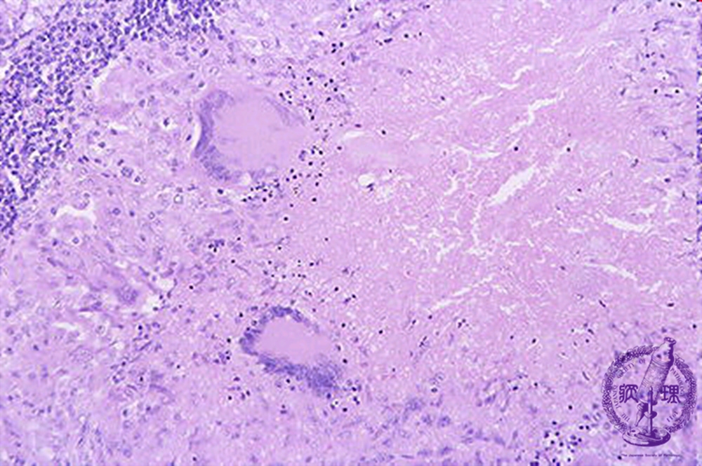
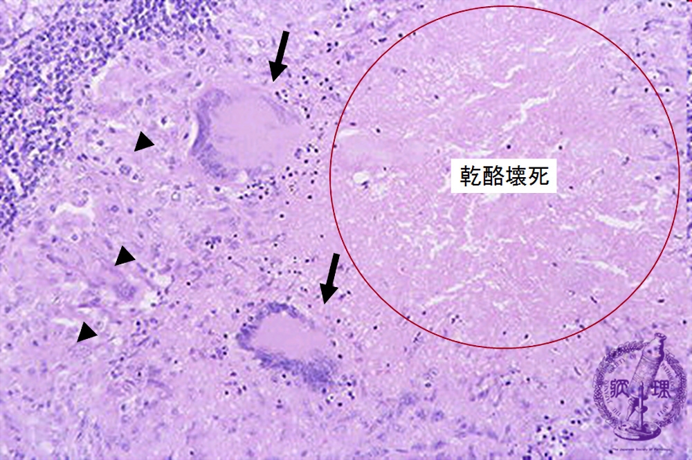
Microscopic image (H&E high power view): In the vicinity of caseous necrosis (circle) are “epithelioid” histiocytes (arrow head) which resemble epithelial cells on account of their round nuclei and abundant pink cytoplasm. The latter cells fuse to form Langerhans giant cells (arrow) with multiple, eccentrically situated nuclei. These can also be seen scattered throughout.
Click the image to see the enlarged image.