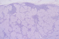


- 2.Lymph node
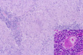
- ★(3)Sarcoidosis
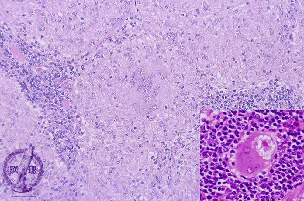
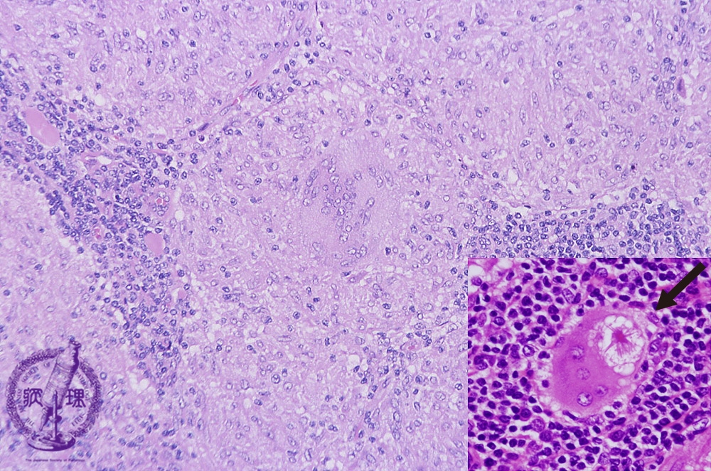
Microscopic image (H&E high power view): Again, epithelioid histiocytes with round nuclei and abundant cytoplasm are seen. Some areas share a strong histologic resemblance to the lesions seen in tuberculosis. However, the lack of necrosis is an important diagnostic point which helps to differentiate sarcoidosis. Langerhans giant cells, again seen here, are a constant feature but their prominence and numbers will vary. An “asteroid” body is also present (arrow at lower right).
Click the image to see the enlarged image.